Groin Muscle Anatomy - Groin - Atlas of Human Anatomy - Centralx : These are the muscles on the inner side of the thigh.. The groin region consists of ligaments, tendons, muscles and fascia all of which attach to the pubic bone. Groin is made of multiple ligaments, muscles, and tendons which fuse together in the pubic bone. Medically, the groin is the junction between the abdomen and thigh. The movement at the joint depends on the anatomy of the joint and its axes of movement. The adductor longus and gracilis both originate form from the pubic bone.
The inguinal ligament is an important connective tissue structure in the inguinal, or groin, region of the human body. The groin region is subdivided into two distinct anatomic areas: The adductor longus and gracilis both originate form from the pubic bone. The groin is the area in the body where the upper thighs meet the lowest part of the abdomen. Hip anatomy, function and common problems front view of the hip joint bones.
Open Inguinal Hernia Repair | Basicmedical Key from basicmedicalkey.com It lies between the chest and the pelvis, holding many of the body's organs. Hip adductor muscles together make up the groin area. Hip anatomy, function and common problems front view of the hip joint bones. Without a clear clinical/pathological diagnosis, the subsequent management of chronic groin pain is difficult. The hip muscles include pelvic and groin muscles. The groin is the area that lies between the abdomen (stomach) and thighs. The gluteus medius, gluteus minimus, piriformis, tensor fasciae latae on the outside. Here we explain the symptoms, causes, treatment, and exercises for a groin muscle strain.
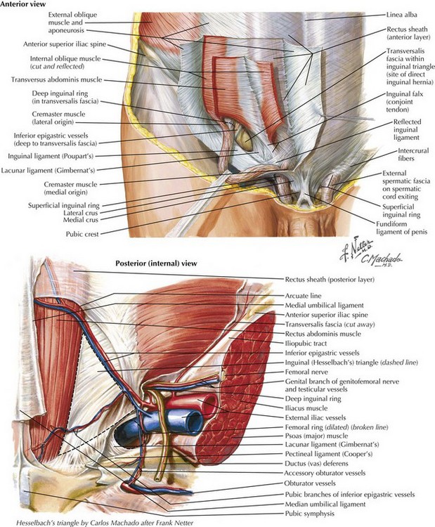
The only openings in the wall are small tunnels called the inguinal and femoral canals.
Often groin strain occurs in the area of inguinal ligament. These are the muscles on the inner side of the thigh. Anatomy of the abdomen and groin. Hip & groin muscles the main hip & groin muscles consist of the iliopsoas, pectineus, rectus femoris, and sartorius at the front. The shoulder muscles are responsible for maintaining the widest range of motion of any joint in your body. Included in this group are the adductor longus, adductor brevis, adductor magnus, pectineus, and gracilis muscles. The lower leg muscles are essential bodily structures. Anatomy of the groin superficial structures of the groin the fascia lata is the deep fascia of the thigh and encloses the muscles and forms the outer limit of the fascial compartments. We'll discuss the function and anatomy. We think this is the most useful anatomy picture that you need. The groin region is subdivided into two distinct anatomic areas: Posterior surface anatomy with muscles that. Without a clear clinical/pathological diagnosis, the subsequent management of chronic groin pain is difficult.
Inferior surface anatomy with underlying pelvis. The groin is the area in the body where the upper thighs meet the lowest part of the abdomen. It supports soft tissues in the groin as well as the external abdominal oblique muscle. Here we explain the symptoms, causes, treatment, and exercises for a groin muscle strain. In human anatomy, the groin is the junctional area between the abdomen and the thigh on either side of the pubic bone.
Groin Muscle Anatomy / Athletic Pubalgia And Sports Hernia ... from lh5.googleusercontent.com The groin is the area that lies between the abdomen (stomach) and thighs. Groin is made of multiple ligaments, muscles, and tendons which fuse together in the pubic bone. Hip adductor muscles together make up the groin area. Posterior surface anatomy with sciatic. Hip & groin muscles the main hip & groin muscles consist of the iliopsoas, pectineus, rectus femoris, and sartorius at the front. The groin region is subdivided into two distinct anatomic areas: We'll discuss the function and anatomy. Groin pain might be worsened by continued use of the injured area.
Anatomy of the groin superficial structures of the groin the fascia lata is the deep fascia of the thigh and encloses the muscles and forms the outer limit of the fascial compartments.
The groin region is subdivided into two distinct anatomic areas: Here we explain the symptoms, causes, treatment, and exercises for a groin muscle strain. We hope this picture groin region anatomy diagram can help you study and research. Groin is made of multiple ligaments, muscles, and tendons which fuse together in the pubic bone. A groin pull is an injury to the muscles (a muscle strain) of the inner thigh. The movement at the joint depends on the anatomy of the joint and its axes of movement. These are the muscles on the inner side of the thigh. Often groin strain occurs in the area of inguinal ligament. Marcella cited as part of the groin. When a teacher talks about moving the groin, they often. A sudden sharp pain is felt which can range from a mild to very severe. Inferior surface anatomy with underlying pelvis. Here we explain the hip and groin muscles, their actions and exercises.
Often groin strain occurs in the area of inguinal ligament. Anatomy of the abdomen and groin. The gluteus medius, gluteus minimus, piriformis, tensor fasciae latae on the outside. Posterior surface anatomy with muscles that. Groin pain might occur immediately after an injury, or pain might come on gradually over a period of weeks or even months.
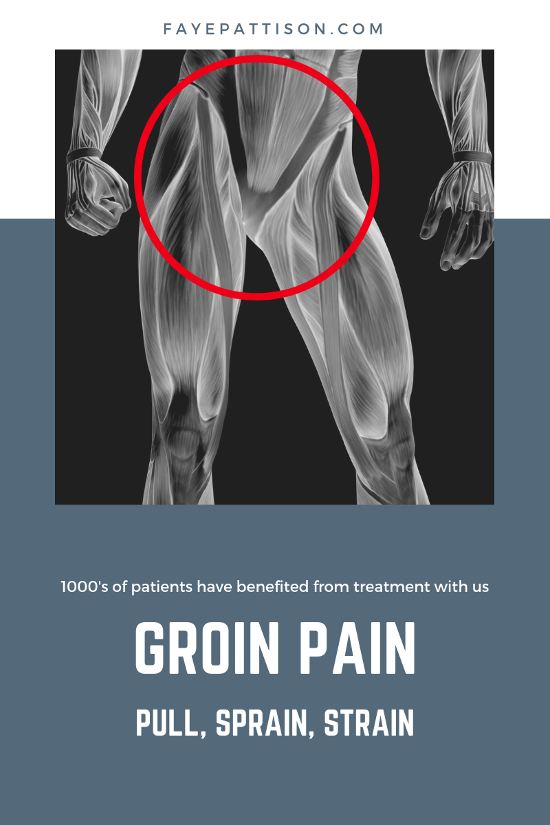
Pulled Groin Muscle Anatomy / Groin Strain Physiopedia ... from fayepattison.com The hip muscles include pelvic and groin muscles. The groin muscles are a group of muscles situated high on the leg in the inner thigh. The movement at the joint depends on the anatomy of the joint and its axes of movement. A groin strain is a tear of the adductor muscles on the inside of the thigh. We think this is the most useful anatomy picture that you need. We'll discuss the function and anatomy. The adductor longus and gracilis both originate form from the pubic bone. Anatomynote.com found groin region anatomy diagram from plenty of anatomical pictures on the internet.
The belly (abdomen) is the largest space (cavity) in the body.
These include the liver, stomach, and intestines. Groin pain might be worsened by continued use of the injured area. A sudden sharp pain is felt which can range from a mild to very severe. It is often referred to as a 'pulled groin muscle', or a 'groin pull'. It is also flexible enough to prevent injury and a. The gluteus medius, gluteus minimus, piriformis, tensor fasciae latae on the outside. The only openings in the wall are small tunnels called the inguinal and femoral canals. Anatomynote.com found groin region anatomy diagram from plenty of anatomical pictures on the internet. This is also known as the medial compartment of the thigh. The inguinal ligament is a narrow band of dense regular fibrous connective tissue in the pelvic region of the body. We'll discuss the function and anatomy. The groin region consists of ligaments, tendons, muscles and fascia all of which attach to the pubic bone. It lies between the chest and the pelvis, holding many of the body's organs.